Ji-jun Liu1,
Xin-wen Wang1,
Shu-fang Wu2,
Qi-ning Wu1,
Ding-jun Hao1 ![]()
For correspondence:- Ding-jun Hao Email: haodingjun@hotmail.com Tel:+862987800002
Received: 18 January 2016 Accepted: 6 July 2016 Published: 30 August 2016
Citation: Liu J, Wang X, Wu S, Wu Q, Hao D. Salvia miltiorrhiza aqueous root extract plays an important role in improving locomotor activity in rats with spinal cord injury. Trop J Pharm Res 2016; 15(8):1667-1672 doi: 10.4314/tjpr.v15i8.11
© 2016 The authors.
This is an Open Access article that uses a funding model which does not charge readers or their institutions for access and distributed under the terms of the Creative Commons Attribution License (http://creativecommons.org/licenses/by/4.0) and the Budapest Open Access Initiative (http://www.budapestopenaccessinitiative.org/read), which permit unrestricted use, distribution, and reproduction in any medium, provided the original work is properly credited..
Purpose: To investigate the activity of the aqueous root extract of Salvia miltiorrhiza (S. miltiorrhiza) (Lamiaceae), collected from Anhui Province, China, for the treatment of spinal cord injury (SCI) in Sprague-Dawley (SD) rats.
Methods: In total, 30 adult rats were selected and divided into three groups; normal control, untreated and treated. Aqueous root extract of S. miltiorrhiza was introduced intraperitoneally to the treated group. Basso, Beattie and Bresnahan rating scale (BBB) was used to evaluate improvement in locomotor activity after SCI. Total RNA was extracted from tissue sections using Sepasol (NacalaiTesque) and RNA samples were reverse-transcribed using M-MLV reverse transcriptase. BioSense SC-810 Gel Documentation System and Gel-Pro 3.1 software were employed for the analysis of band intensity.
Results: A significant reduction in SCI cavity area was observed in the S. miltiorrhiza extract-treated group, relative to the untreated group, after 11 days (0.10 ± 0.05 mm2 treated vs. 0.30 ± 0.01 mm2 untreated). Treatment with root extract also improved the BBB scores; the treated group scored 15, compared to a score of 8 for the untreated group. In addition, the degradation of neurons at the site of injury in the spinal cord was reduced in the treated group compared to the untreated group. Treatment with S. miltiorrhiza aqueous root extract also significantly increased the ex
Conclusion: These data suggest that, in addition to other pharmacological activities, S. miltiorrhiza extract has therapeutic potential for the treatment of neuronal degeneration following SCI.
Introduction
Spinal cord injury (SCI), which is primarily caused by traffic accidents and falls from high altitude, is a frequently encountered global health problem [1]. The pathology of SCI can be divided into two stages, primary and secondary injury, involving (1) compression of spinal cord tissues and (2) apoptosis of cells at the site of lesions, respectively [2,3]. The permanent loss of neurological functions and the associated life-threatening complications make SCI a major clinical issue across the globe [4]. SCI involves loss of either partial or complete neuronal sensory and motor functions, because of the failure of neuronal regeneration. Axonal regeneration is inhibited by autonomous cellular factors [5,6], astroglial scarring [7,8] and by the accumulation of central nervous system (CNS) myelin [8,9].
There are cells in the CNS that produce platelet-derived growth factor (PDGF), which has been demonstrated to bind to NMDA receptors [10]. It is reported that PDGF plays a vital role in recovery following CNS injury [10]. The mechanism of action of PDGF involves activation of tyrosine kinase receptors, including PDGF-αR and PDGF-βR. PDGF has also been identified to promote the growth of axons and to maintain the integrity of neurons [11]. PDGF isoforms induce mitosis in glial cells, thereby conferring a protective effect on the brain.
Natural products, either in the form of extracts or isolated constituents, have been used for the treatment of various disorders. Extracts from plants like Lycium barbarum (Solanaceae) and S. miltiorrhiza root (Lamiaceae) have been shown to possess neuroprotective activities [12,13]. It has been reported that the alcoholic root and rhizome extract of S. miltiorrhiza (Lamiaceae) protects the eyes of elderly people and provides relief from strain and pain [14,15]. The present study was designed to investigate the effect of S. miltiorrhiza root extract on rats with SCI. It was observed that S. miltiorrhiza (Lamiaceae) root extract improved locomotor activity, indicating that it may be a promising treatment option for SCI.
Methods
Animals
Thirty adult (8 weeks of age) Sprague-Dawley (SD) rats were obtained from Guangdong Medical Laboratory Animal Co. (Guangdong, China). The animals were acclimatized to the laboratory atmosphere for 1 week prior to commencement of the experiment, and were housed under 12 h dark/light cycle conditions with free access to water. Experimental procedures were carried out according to the guidelines approved by the Animal care and use committee of Xi'an Jiaotong University (approval no. 2014/10C-25). The studies were carried out in accordance with the Directive Office of Laboratory Animal Welfare, National Institutes of Health (OLAW/NIH 2002) on the guidelines for care and safe use of laboratory animals for scientific purposes [16].
Plant extract
Initially, the plant samples were collected from Gunagdong, Sichuan, and Shangdong Provinces and all specimens were examined, identified and authenticated by Dr. H.W. Wang, Plant Taxonomist, China Agricultural University. The specimens were registered and the voucher specimen (no. HKLPR/IBA/CAS/consult/-2014-03/438/161) deposited in the Herbarium of the Key Laboratory of Plant Resources, Institute of Botany, Chinese Academy of Sciences.
S. miltiorrhiza (Lamiaceae) plant root was collected, washed with distilled water, dried and ground to form a fine powder. The powder was added to a mixture of distilled water and methanol (1:1 ratio) and macerated for 2 days, after which the solvent mixture was evaporated and filtered through Whatman paper to obtain a pure extract.
Reagents and chemicals
Dimethyl sulphoxide (DMSO) and 3,3'-diaminobenzidine hydrochloride (DAB) were purchased from Sigma Co. (St. Louis, MO, USA). Antibodies for PDGF-B and biotinylated goat anti-rabbit secondary antibody were obtained from Santa Cruz Biotechnology, Inc. (Santa Cruz, CA, USA).
Treatment strategy
The rats were divided randomly into three groups (normal control, untreated and treated) of 10 animals each. The animals in the untreated and treated groups were anesthetized by intra peritoneal injection of sodium pentobarbital (50 mg/kg), before exposure of the spinal cord. A contusion injury was carefully made in the spinal cord of rats in both groups using a weight-drop device [17]. The incision was closed by sterile suturing and the animals were kept in their respective marked cages under sterilized conditions.
Animals in the treated group were injected with 10 µM doses of S. miltiorrhiza root extract every other day for 10 days. Animals in the untreated and normal control groups were injected with the same volume (5 mL/kg) of normal saline. On day 11, the animals were placed on the floor and their locomotor ability was monitored for 10 min [18]. Locomotor capacity was scored based on the Basso, Beattie and Bresnahan rating scale (BBB).
Examination of pathological changes [10]
On day 11, four animals from each group were anesthetized and a surgical incision was performed to open the thoracic cavity. A segment of the thoracic spinal cord from T6 to T14 was removed from each animal, washed in normal saline and fixed in 4 % paraformaldehyde in 0.1 M phosphate-buffered saline (PBS) for 5 h. The tissue sections were rinsed twice in PBS and then incubated for 45 min with 1 % bovine serum albumin (BSA). The sections were then incubated with primary antibodies (anti-PDGF-B primary antibody) in BSA for 12 h. Following incubation, the sections were washed again with PBS and incubated with biotinylated goat anti-rabbit secondary antibody.
The sections were then treated with avidin-horse radish peroxidase (HRP) solution. Visualization was achieved using DAB and hydrogen peroxide dissolved in PBS. The paraffin-embedded sections were heated in xylene, and hydrated in a gradient of ethyl alcohol. Finally, the sections were mounted onto poly-L-lysine-coated slides using Permount™. The lesion sites were examined under a microscope using H&E staining.
Analysis of PDGF-B mRNA expression [18]
Total RNA was extracted from tissue sections using Sepasol (NacalaiTesque) according to the manufacturer’s protocol. Moloney-murine leukemia virus (M-MLV) reverse transcriptase (Life Technologies Japan Ltd., Tokyo, Japan) was used to reverse-transcribe the RNA samples, using a thermal cycler (Takara PCR Thermal Cycler S; Takara Bio Inc., Shiga, Japan). The mRNA expression levels were evaluated using a real-time quantitative RT-PCR assay and a Light Cycler System (Roche Diagnostics, Mannheim, Germany).
Separation of the PCR products was performed using electrophoresis on a 2.0 % agarose gel. A BioSense SC-810 Gel Documentation System (Shanghai Bio-Tech Co., Ltd., Shanghai, China) and Gel-Pro software (ver. 3.1; Media Cybernetics, Inc., Bethesda, MD, USA) were employed for the analysis of the intensity of the bands.
Statistical analysis
All of the experiments were performed in triplicate and the data are presented as mean ± standard deviation (SD). Statistical analysis of the data was performed using SPSS for Windows software (ver. 16.0; SPSS Inc., Chicago, IL, USA).
Statistically significant differences were identified using one-way analysis of variance (ANOVA). Differences were considered significant at p < 0.05.
Results
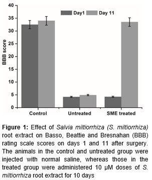
S. miltiorrhiza root extract increased the BBB score
The animals belonging to the normal control, untreated and treated groups were examined for locomotor activity on day 11 post-surgery. It was observed that animals in the untreated and treated groups suffered from hind limb paralysis and were unable to walk normally in the initial days after the saline or root extract injection regimen was initiated, whereas animals in the normal control group walked normally. However, treatment with S. miltiorrhiza root extract improved recovery from hind limb paralysis; animals in the treated group started to walk normally after day 10. The BBB score in the S. miltiorrhiza root extract-treated group improved, reaching 15 points, compared to the 8 points scored by the untreated group of rats (). These findings indicate that S. miltiorrhiza root extract has a therapeutic effect on rats following SCI (p < 0.01).
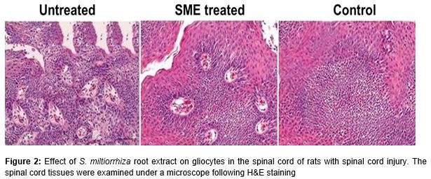
The results from H&E staining revealed that the spinal cord tissues of the untreated group of animals featured vacuolar regions, reduced numbers of gliocytes and neuronal apoptosis. However, treatment of the rats with S. miltiorrhiza root extract prevented both the formation of vacuoles and neuronal apoptosis. The animals in the treatment group were found to possess large numbers of gliocytes in the spinal cord at the site of injury ().
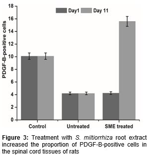
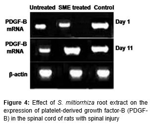
S. miltiorrhiza root extract increases the expression of PDGF-B
DAB staining revealed that treatment of the animals with S. miltiorrhiza root extract induced an increase in the proportion of PDGF-B-positive cells. Compared to the untreated group, animals in the S. miltiorrhiza root extract-treated group showed significantly higher numbers of PDGF-B-positive cells on day 11 after surgery (). S. miltiorrhiza root extract also resulted in a reduction in the vacuolar region of the spinal cord following injury. Analysis of the expression of mRNA corresponding to PDGF-B revealed a marked increase in the treated group compared to the untreated group (). The expression of mRNA corresponding to PDGF-B in the treated group was 7-fold higher compared to the untreated group.
Discussion
Extracts of plants such as Lycium barbarum and S. miltiorrhiza have been shown to possess promising neuroprotective activities [12,13]. In the present study, S. miltiorrhiza root extract was shown to be beneficial for the treatment of rats with SCI. S. miltiorrhiza root extract induced a significant improvement in recovery compared to untreated rats. Rats in the treatment group showed significantly higher BBB scores and reduced neuronal apoptosis 11 days after surgery. Axons were also found to show regeneration in animals treated with S. miltiorrhiza root extract.
The PDGF, which has been demonstrated to bind to NMDA receptors, is secreted by cells in the CNS [10]. It is reported that PDGF plays a vital role in recovery following CNS injury [10]. The mechanism of action of PDGF involves activation of tyrosine kinase receptors, including PDGF-αR and PDGF-βR. PDGF has also been shown to promote the growth of axons and to maintain the integrity of neurons [11]. PDGF isoforms induce mitosis in glial cells, thereby affecting a protective effect on the brain.
It is reported that PDGF protects dopaminergic neurons from toxicity induced by Tat proteins. Increasing PDGF expression may therefore be a promising therapeutic strategy for SCI. The results from the present study revealed that S. miltiorrhiza root extract enhances the expression of PDGF-B 10 days after surgery. The improvement in recovery of rats treated with S. miltiorrhiza root extract following SCI may therefore be associated with promotion of PDGF-B expression. Reports indicate that the proportion of PDGF-positive cells in the spinal cord of rats is significantly higher following SCI [17,21]. PDGF-B plays an important role in the protection of neurons by inhibiting glutamate transporters in the cell membrane. Furthermore, enhancement of the PDGF-B/receptor signal pathway may rescue neonatal brains at risk of hypoxic-ischemic injury [22].
Thus, we conclude that the root extract of S. miltiorrhiza has the capacity to improve recovery from SCI in rats, via a mechanism involving promotion of PDGF-B expression. Therefore, Salvia miltiorrhiza extract may be of potential therapeutic use for treatment of SCI.
Conclusion
The findings of the present study demonstrate that the root extract of S. miltiorrhiza significantly improves recovery, as indicated by increase in locomotor activity, following SCI injury in a rat model. The results also suggest that S. miltiorrhiza root extract has therapeutic potentials for the treatment of neuronal degeneration associated with spinal cord injuries.
Declarations
Acknowledgement
References
Archives


News Updates